

Which Medical Term Means A Small, Circumscribed Elevation On The Skin?
| Skin condition | |
|---|---|
| Other names | Cutaneous status |
 | |
| 3D medical illustration showing major layers of pare | |
| Specialty | Dermatology |
| Causes | Sun exposure, vitamin deficiencies, substance usage, poor hygiene, cancers |
A skin condition, also known as cutaneous condition, is any medical condition that affects the integumentary organization—the organ system that encloses the body and includes skin, nails, and related muscle and glands.[1] The major part of this system is equally a barrier against the external surroundings.[2]
Conditions of the human integumentary system establish a broad spectrum of diseases, also known as dermatoses, as well as many nonpathologic states (similar, in certain circumstances, melanonychia and racquet nails).[3] [4] While merely a small number of skin diseases account for nigh visits to the medico, thousands of skin conditions take been described.[v] Nomenclature of these conditions oft presents many nosological challenges, since underlying causes and pathogenetics are often non known.[6] [7] Therefore, most electric current textbooks present a nomenclature based on location (for instance, conditions of the mucous membrane), morphology (chronic blistering conditions), crusade (skin weather resulting from concrete factors), and then on.[8] [9]
Clinically, the diagnosis of any particular skin status begins by gathering pertinent data of the presenting skin lesion(s), including: location (arms, caput, legs); symptoms (pruritus, hurting); duration (astute or chronic); arrangement (solitary, generalized, annular, linear); morphology (macules, papules, vesicles); and colour (red).[10] Some diagnoses may also require a skin biopsy which yields histologic data[eleven] [12] that can exist correlated with the clinical presentation and any laboratory information.[xiii] [fourteen] The introduction of cutaneous ultrasound has immune the detection of cutaneous tumors, inflammatory processes, and skin diseases.[15]
Layer of pare involved [edit]
The skin weighs an boilerplate of iv kg (8.8 lb), covers an area of 2 one thousand2 (22 sq ft), and is fabricated of three distinct layers: the epidermis, dermis, and subcutaneous tissue.[i] The two primary types of human peel are glabrous skin, the nonhairy pare on the palms and soles (also referred to as the "palmoplantar" surfaces), and hair-bearing skin.[16] Within the latter type, hairs in structures called pilosebaceous units have a hair follicle, sebaceous gland, and associated arrector pili muscle.[17] In the embryo, the epidermis, hair, and glands are from the ectoderm, which is chemically influenced by the underlying mesoderm that forms the dermis and subcutaneous tissues.[18] [xix] [xx]
Epidermis [edit]
The epidermis is the most superficial layer of skin, a squamous epithelium with several strata: the stratum corneum, stratum lucidum, stratum granulosum, stratum spinosum, and stratum basale.[21] Nourishment is provided to these layers via diffusion from the dermis, since the epidermis is without direct claret supply.[22] The epidermis contains 4 prison cell types: keratinocytes, melanocytes, Langerhans cells, and Merkel cells. Of these, keratinocytes are the major component, constituting roughly 95% of the epidermis.[16] This stratified squamous epithelium is maintained by cell segmentation within the stratum basale, in which differentiating cells slowly displace outwards through the stratum spinosum to the stratum corneum, where cells are continually shed from the surface.[16] In normal skin, the rate of production equals the charge per unit of loss; about two weeks are needed for a cell to drift from the basal cell layer to the top of the granular cell layer, and an boosted ii weeks to cross the stratum corneum.[23]
Dermis [edit]
The dermis is the layer of skin between the epidermis and subcutaneous tissue, and comprises ii sections, the papillary dermis and the reticular dermis.[24] The superficial papillary dermis interdigitates with the overlying rete ridges of the epidermis, betwixt which the ii layers interact through the basement membrane zone.[24] Structural components of the dermis are collagen, elastic fibers, and ground substance too called extra fibrillar matrix.[24] Within these components are the pilosebaceous units, arrector pili muscles, and the eccrine and apocrine glands.[21] The dermis contains 2 vascular networks that run parallel to the skin surface—one superficial and one deep plexus—which are continued by vertical communicating vessels.[21] [25] The role of blood vessels within the dermis is fourfold: to supply nutrition, to regulate temperature, to attune inflammation, and to participate in wound healing.[26] [27]
Subcutaneous tissue [edit]
The subcutaneous tissue is a layer of fat betwixt the dermis and underlying fascia.[5] This tissue may be further divided into 2 components, the actual fatty layer, or panniculus adiposus, and a deeper vestigial layer of muscle, the panniculus carnosus.[xvi] The master cellular component of this tissue is the adipocyte, or fat cell.[5] The structure of this tissue is composed of septal (i.e. linear strands) and lobular compartments, which differ in microscopic appearance.[21] Functionally, the subcutaneous fat insulates the body, absorbs trauma, and serves equally a reserve energy source.[5]
Diseases of the pare [edit]
Diseases of the peel include skin infections and skin neoplasms (including skin cancer).[28]
History [edit]
In 1572, Geronimo Mercuriali of Forlì, Italia, completed De morbis cutaneis ('On the diseases of the pare'). Information technology is considered the outset scientific work dedicated to dermatology.
Diagnoses [edit]
The physical examination of the peel and its appendages, as well as the mucous membranes, forms the cornerstone of an accurate diagnosis of cutaneous conditions.[29] Most of these weather present with cutaneous surface changes termed "lesions," which have more or less distinct characteristics.[30] Frequently proper examination will atomic number 82 the physician to obtain advisable historical information and/or laboratory tests that are able to ostend the diagnosis.[29] Upon test, the important clinical observations are the (1) morphology, (ii) configuration, and (iii) distribution of the lesion(s).[29] With regard to morphology, the initial lesion that characterizes a condition is known as the "master lesion", and identification of such a lesions is the most important aspect of the cutaneous examination.[thirty] Over time, these chief lesions may continue to develop or be modified by regression or trauma, producing "secondary lesions".[one] However, with that existence stated, the lack of standardization of basic dermatologic terminology has been one of the principal barriers to successful communication among physicians in describing cutaneous findings.[21] All the same, there are some commonly accepted terms used to draw the macroscopic morphology, configuration, and distribution of skin lesions, which are listed below.[30]
Lesions [edit]
Primary lesions [edit]

Chigger bites on human being peel showing characteristic welts
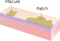
Macule and patch
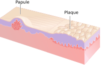
Papule and plaque

Nodules

Vesicles and bulla

Fissures, erosions and ulcers
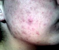
A pustule on the cheek

Relative incidence of skin cysts
- Macule: A macule is a modify in surface color, without top or depression, and then nonpalpable, well or ill-defined,[10] variously sized, but generally considered less than either v[10] or 10 mm in bore at the widest point.[30]
- Patch: A patch is a big macule equal to or greater than either 5 or 10 mm across,[30] depending on ane's definition of a macule.[1] Patches may accept some subtle surface change, such equally a fine calibration or wrinkling, but although the consistency of the surface is inverse, the lesion itself is not palpable.[29]
- Papule: A papule is a circumscribed, solid height of skin, varying in size from less than either 5[ten] or ten mm in diameter at the widest betoken.[30]
- Plaque: A plaque has been described as a broad papule, or confluence of papules equal to or greater than ten mm,[30] or alternatively as an elevated, plateau-like lesion that is greater in its bore than in its depth.[29]
- Nodule: A nodule is morphologically similar to a papule in that it is also a palpable spherical lesion less than ten mm in bore. However, it is differentiated by being centered deeper in the dermis or subcutis.
- Tumor: Similar to a nodule, but it is larger than 10 mm in diameter.[ citation needed ]
- Vesicle: A vesicle is minor blister,[31] a confining, epidermal meridian generally considered less than either 5[10] or ten mm in diameter at the widest indicate.[xxx]
- Bulla: A bulla is a big blister,[31] a rounded or irregularly shaped blister equal to or greater than either 5[10] or 10 mm,[30] depending on one's definition of a vesicle.[i]
- Pustule: A pustule is a small-scale top of the skin usually consisting of necrotic inflammatory cells.[xxx]
- Cyst: A cyst is an epithelial-lined cavity. [10]
- Wheal: A wheal is a rounded or flat-topped, pale ruby papule or plaque that is characteristically evanescent, disappearing within 24 to 48 hours. The temporary raised skin on the site of a properly delivered intradermal (ID) injection is likewise chosen a welt, with the ID injection process itself oftentimes referred to every bit simply "raising a wheal" in medical texts.[10]
- Welts: Welts occur as a event of blunt force existence applied to the torso with elongated objects without sharp edges.
- Telangiectasia: A telangiectasia represents an enlargement of superficial blood vessels to the point of being visible.[29]
- Burrow: A burrow appears every bit a slightly elevated, grayish, tortuous line in the skin, and is acquired by burrowing organisms.[29] [thirty]
Secondary lesions [edit]
- Scale: Dry or greasy laminated masses of keratin,[xxx] they correspond thickened stratum corneum.[29]
- Chaff: Dried sebum normally mixed with epithelial and sometimes bacterial debris[10]
- Lichenification: Epidermal thickening characterized by visible and palpable thickening of the skin with accentuated pare markings[1]
- Erosion: An erosion is a aperture of the pare exhibiting incomplete loss of the epidermis,[32] a lesion that is moist, confining, and usually depressed.[21] [33]
- Excoriation: A punctate or linear abrasion produced by mechanical means (often scratching), usually involving only the epidermis, but commonly reaching the papillary dermis[30] [33]
- Ulcer: An ulcer is a aperture of the skin exhibiting complete loss of the epidermis and often portions of the dermis.[32] [33]
- Cleft is a lesion in the skin that is usually narrow but deep.[29] [33]
- Induration is dermal thickening causing the cutaneous surface to feel thicker and firmer.[29]
- Cloudburst refers to a loss of skin, and can be epidermal, dermal, or subcutaneous.[30] With epidermal atrophy, the skin appears sparse, translucent, and wrinkled.[29] Dermal or subcutaneous cloudburst is represented by depression of the skin.[29]
- Maceration: softening and turning white of the skin due to beingness consistently moisture.
- Umbilication is formation of a depression at the pinnacle of a papule, vesicle, or pustule.[34]
- Phyma: A tubercle on any external part of the body, such equally in phymatous rosacea
Configuration [edit]
"Configuration" refers to how lesions are locally grouped ("organized"), which contrasts with how they are distributed (see adjacent section).
- Agminate: in clusters
- Annular or circinate: band-shaped
- Arciform or arcuate: arc-shaped
- Digitate: with finger-similar projections
- Discoid or nummular: circular or disc-shaped
- Figurate: with a detail shape
- Guttate: resembling drops
- Gyrate: coiled or spiral-shaped
- Herpetiform: resembling herpes
- Linear
- Mammillated: with rounded, breast-similar projections
- Reticular or reticulated: resembling a net
- Serpiginous: with a wavy border
- Stellate: star-shaped
- Targetoid: resembling a bullseye
- Verrucous or Verruciform: wart-like
Distribution [edit]
"Distribution" refers to how lesions are localized. They may be confined to a single area (a patch) or may be in several places. Some distributions correlate with the means by which a given area becomes affected. For case, contact dermatitis correlates with locations where allergen has elicited an allergic immune response. Varicella zoster virus is known to recur (afterwards its initial presentation as chicken pox) as herpes zoster ("shingles"). Chicken pox appears well-nigh everywhere on the body, only herpes zoster tends to follow 1 or two dermatomes; for example, the eruptions may appear forth the bra line, on either or both sides of the patient.[ citation needed ]
- Generalized
- Symmetric: one side mirrors the other
- Flexural: on the front of the fingers
- Extensor: on the back of the fingers
- Intertriginous: in an area where 2 peel areas may impact or rub together
- Morbilliform: resembling measles
- Palmoplantar: on the palm of the hand or bottom of the foot
- Periorificial: around an orifice such as the rima oris
- Periungual/subungual: around or under a fingernail or toenail
- Blaschkoid: following the path of Blaschko'south lines in the skin
- Photodistributed: in places where sunlight reaches
- Zosteriform or dermatomal: associated with a particular nervus
[edit]
- Collarette
- Comedo
- Confluent
- Eczema (a blazon of dermatitis)
- Evanescent (lasting less than 24 hours)
- Granuloma
- Livedo
- Purpura
- Erythema (redness)
- Horn (a cell blazon)
- Poikiloderma
Histopathology [edit]
- Hyperkeratosis
- Parakeratosis
- Hypergranulosis
- Acanthosis
- Papillomatosis
- Dyskeratosis
- Acantholysis
- Spongiosis
- Hydropic swelling
- Exocytosis
- Vacuolization
- Erosion
- Ulceration
- Lentiginous
See besides [edit]
- Wound, an injury which damages the epidermis.
References [edit]
- ^ a b c d due east f Miller, Jeffrey H.; Marks, James G. (2006). Lookingbill and Marks' Principles of Dermatology. Saunders. ISBNi-4160-3185-5.
- ^ Lippens, S; Hoste, E; Vandenabeele, P; Agostinis, P; Declercq, W (April 2009). "Cell death in the skin". Apoptosis. 14 (4): 549–69. doi:10.1007/s10495-009-0324-z. PMID 19221876. S2CID 13058619.
- ^ King, L.Southward. (1954). "What Is Disease?". Philosophy of Science. 21 (3): 193–203. doi:x.1086/287343. S2CID 120875348.
- ^ Bluefarb, Samuel Yard. (1984). Dermatology . Upjohn Co. ISBN0-89501-004-half-dozen.
- ^ a b c d Lynch, Peter J. (1994). Dermatology. Williams & Wilkins. ISBN0-683-05252-7.
- ^ Tilles G, Wallach D (1989). "[The history of nosology in dermatology]". Ann Dermatol Venereol (in French). 116 (1): 9–26. PMID 2653160.
- ^ Lambert WC, Everett MA (Oct 1981). "The nosology of parapsoriasis". J. Am. Acad. Dermatol. 5 (4): 373–95. doi:10.1016/S0190-9622(81)70100-two. PMID 7026622.
- ^ Jackson R (1977). "Historical outline of attempts to classify skin diseases". Can Med Assoc J. 116 (x): 1165–68. PMC1879511. PMID 324589.
- ^ Copeman PW (February 1995). "The creation of global dermatology". J R Soc Med. 88 (2): 78–84. PMC1295100. PMID 7769599.
- ^ a b c d e f chiliad h i Wolff, Klaus; Johnson, Richard Allen; Suurmond, Richard (2005). Fitzpatrick's Color Atlas and Synopsis of Clinical Dermatology (5th ed.). McGraw-Hill Medical Pub. Sectionalisation. ISBN0-07-144019-four.
- ^ Werner B (August 2009). "[Skin biopsy and its histopathologic analysis: Why? What for? How? Function I]". An Bras Dermatol (in Portuguese). 84 (4): 391–95. doi:10.1590/s0365-05962009000400010. PMID 19851671.
- ^ Werner B (Oct 2009). "[Pare biopsy with histopathologic analysis: why? what for? how? part II]". An Bras Dermatol (in Portuguese). 84 (5): 507–thirteen. doi:10.1590/S0365-05962009000500010. PMID 20098854.
- ^ Xiaowei Xu; Elder, David A.; Rosalie Elenitsas; Johnson, Bernett L.; Murphy, George E. (2008). Lever'southward Histopathology of the Skin. Hagerstwon, Doctor: Lippincott Williams & Wilkins. ISBN978-0-7817-7363-8.
- ^ Weedon's Pare Pathology, 2-Volume Set: Expert Consult – Online and Print. Edinburgh: Churchill Livingstone. 2009. ISBN978-0-7020-3941-6.
- ^ Alfageme, Fernando; Cerezo, Eugenio; Roustan, Gaston (Apr 2015). "Real-Time Elastography in Inflammatory Skin Diseases: A Primer". Ultrasound in Medicine & Biology. 41 (4): S82–S83. doi:10.1016/j.ultrasmedbio.2014.12.341.
- ^ a b c d Burns, Tony; et al. (2006) Rook'south Textbook of Dermatology CD-ROM. Wiley-Blackwell. ISBN 1-4051-3130-half-dozen.
- ^ Paus R, Cotsarelis G (1999). "The biological science of pilus follicles". N Engl J Med. 341 (7): 491–97. doi:10.1056/NEJM199908123410706. PMID 10441606.
- ^ Goldsmith, Lowell A. (1983). Biochemistry and physiology of the peel. Oxford University Press. ISBN0-19-261253-0.
- ^ Fuchs E (Feb 2007). "Scratching the surface of skin development". Nature. 445 (7130): 834–42. Bibcode:2007Natur.445..834F. doi:10.1038/nature05659. PMC2405926. PMID 17314969.
- ^ Fuchs Eastward, Horsley V (Apr 2008). "More 1 way to skin ". Genes Dev. 22 (8): 976–85. doi:10.1101/gad.1645908. PMC2732395. PMID 18413712.
- ^ a b c d east f Wolff, Klaus Dieter; et al. (2008). Fitzpatrick's Dermatology in General Medicine. McGraw-Hill Medical. ISBN978-0-07-146690-5.
- ^ "Skin Anatomy". Medscape. Retrieved 3 June 2013.
- ^ Bolognia, Jean L.; et al. (2007). Dermatology. St. Louis: Mosby. ISBN978-1-4160-2999-one.
- ^ a b c Rapini, Ronald P. (2005). Applied dermatopathology. Elsevier Mosby. ISBN0-323-01198-5.
- ^ Grant-Kels, JM (2007). Color Atlas of Dermatopathology (Dermatology: Clinical & Basic Science). Informa Healthcare. p. 163. ISBN978-0-8493-3794-9.
- ^ Ryan, T (1991). "Cutaneous Apportionment". In Goldsmith, Lowell A (ed.). Physiology, biochemistry, and molecular biology of the skin (2nd ed.). New York: Oxford University Press. p. 1019. ISBN0-19-505612-4.
- ^ Swerlick, RA; Lawley, TJ (Jan 1993). "Role of microvascular endothelial cells in inflammation". J. Invest. Dermatol. 100 (1): 111S–115S. doi:x.1038/jid.1993.33. PMID 8423379.
- ^ Rose, Lewis C. (1998-09-xv). "Recognizing Neoplastic Skin Lesions: A Photo Guide". American Family Physician. 58 (4): 873–84, 887–8. PMID 9767724. Retrieved 3 June 2013.
- ^ a b c d e f g h i j k l Callen, Jeffrey (2000). Color atlas of dermatology. Philadelphia: W.B. Saunders. ISBN0-7216-8256-1.
- ^ a b c d eastward f g h i j k l m n James, William D.; et al. (2006). Andrews' Diseases of the Skin: Clinical Dermatology. Saunders Elsevier. ISBN0-7216-2921-0.
- ^ a b Elsevier, Dorland'south Illustrated Medical Lexicon, Elsevier.
- ^ a b Cotran, Ramzi S.; Kumar, Vinay; Fausto, Nelson; Nelso Fausto; Robbins, Stanley L.; Abbas, Abul 1000. (2005). Robbins and Cotran pathologic basis of disease. St. Louis, Mo: Elsevier Saunders. ISBN0-7216-0187-i.
- ^ a b c d Copstead, Lee-Ellen C.; Diestelmeier, Ruth E.; Diestelmeier, Michael R. (2016-09-03). "Alterations in the Integumentary System". Basicmedical Key . Retrieved 2019-07-01 .
- ^ "Description of Skin Lesions". The Merck Transmission. Retrieved iii June 2013.
External links [edit]

Source: https://en.wikipedia.org/wiki/Skin_condition
Posted by: stoneyoutims.blogspot.com
0 Response to "Which Medical Term Means A Small, Circumscribed Elevation On The Skin?"
Post a Comment